
Salvia divinorum Epling & Játiva, Bot. Mus. Leafl. 20: 75 1962. ;
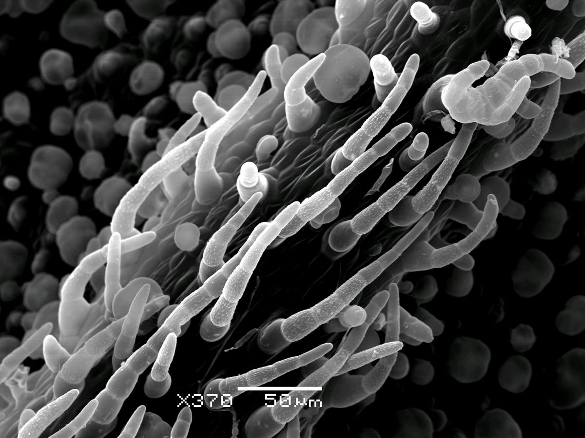
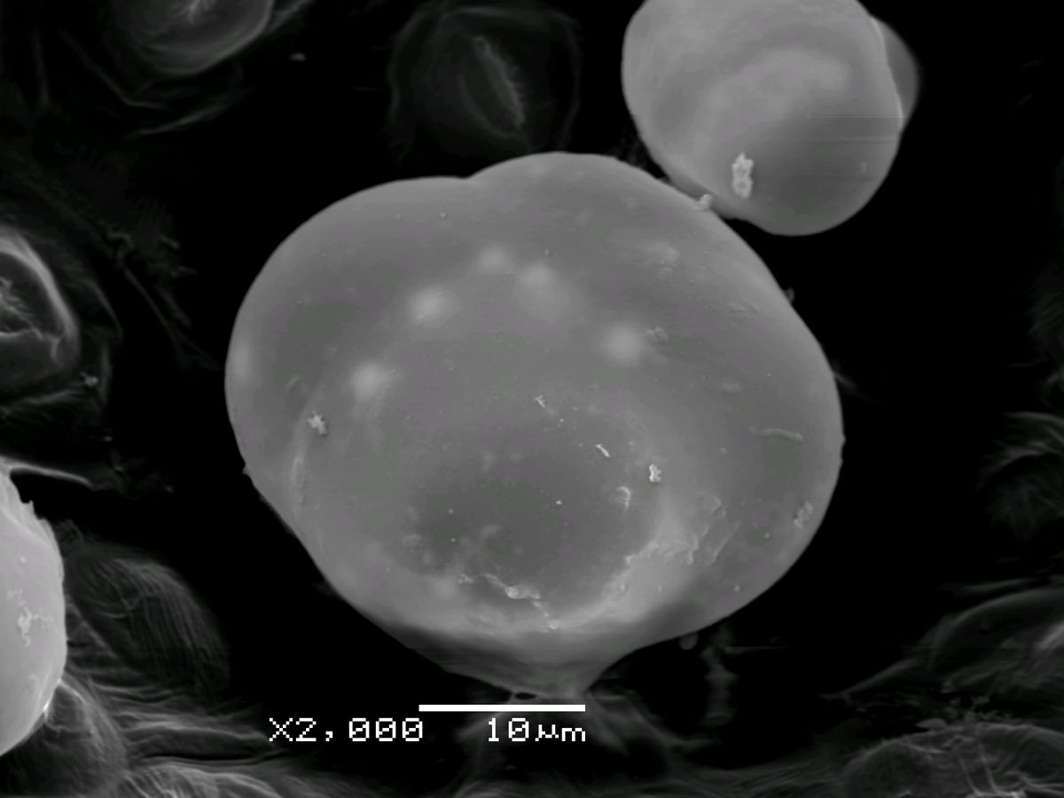
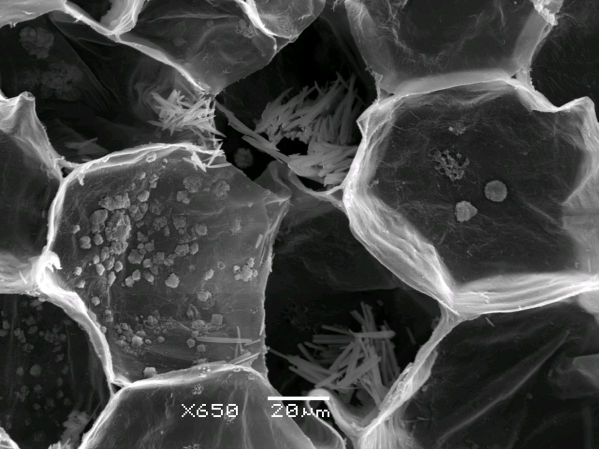
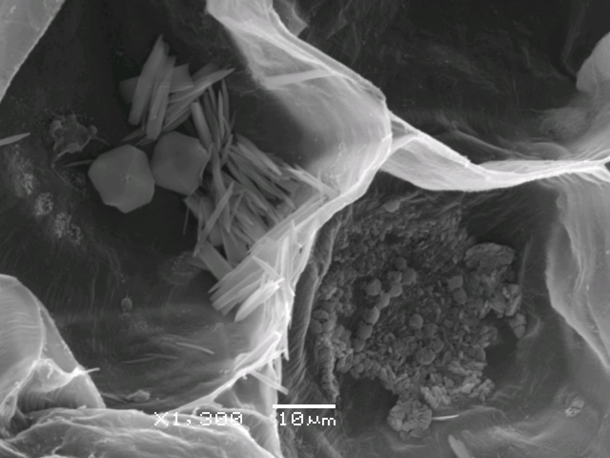
My Flora Picture of the Year 2011: Vijayasankar Raman: Here is yet another b&w picture as ‘my flora picture of the year 2011’. I am not sure if this is suitable for this title, though. But this is my first ever SEM picture 🙂 It was my dream….finally came true! It was a great experience to learn the techniques without a tutor (but now I am helping many…). The picture shows glandular and non-glandular trichomes from leaf of Salvia divinorum, a psychoactive plant endemic to Mexico. I recorded 5 types and 33 sub-types of trichomes (some of them are new reports), from a single leaf, thanks to SEM… This really awesome ooooooo…. this is cool…. unique pictures among all… Best picture ever seen … thanks for showing us the rare beauty and developmental stage which we used to read during PG. We had a SEM in our dept. in Utkal University, but never ever got an opportunity to have a glimpse of it. Even i requested them to see the cell division stage of algae during my phd work, but not permitted again. Thanks a lot again for showing this one. Beauty and wonder at micro-level. Liked this view of Salvia divinorum ! The species name divinorum also means “divine”. It gives a divine feeling to imagine, how on earth such things can be created out of single cell!!! Yes, this plant is called as ‘diviner’s sage’ as it was/is used by the local God men in Mexico to ‘attain divinity’. The active compound Salvinorin-A, that present in the glandular trichomes of the plant, has psychoactive effects. It is believed that this diviner’s sage helps one to attain the state of God. But, since it is highly misused, the consumption and possession have been banned in some countries. Attaching pictures of glandular trichome and cells containing calcium oxalate crystals. A I am so excited … 1: first for you, this will add some new or deeper insights to your ongoing studies…. but of course you knew that…! 2: I am excited for myself and the whole of eflora family… we will get to see the botanical specimen in further details… as and when you share your finds… ================== B: Now, you will be … micromini …. instead of / in addition to me… the micromini epithet was attached to my name by a few friends and colleagues a couple or three decades ago because I was fond of doing TEM and SEM of tumors etc….and ribosome…and mitochondria… ======== C: and this the question section 🙂 the first pic is surface of leaf….. i imagine…. but what are 2 and 3rd pictures of … a fracture of what? is it of a leaf and the rhoboid? structures cell walls… with crystals etc inside? ============== just cant stop smiling…. what a great new dimension to our interaction will this make…. wonderful for all of us… and congratulations for learning the techniques… …, 2nd and third pics show crystals (raphides) inside the cell. I now … and …, you have started confusing me. I can see a single picture, a SEM of leaf surface showing trichomes. I can’t see any second and third picture. Moreover crystals raphides and styloides would be visible in a TEM and not SEM Please correct me if I am wrong. crystals I could tell… Sir he added three more pics later on the same thread 🙂 Please check on the group. I imagine why we cant see raphides under SEM. There has been few studies in India also on Asteraceae I assume by … Should we say First picture is SEM of leaf showing trichomes Second and third picture are TEM of cells showing crystals 4th again seems to be SEM I dont mean to correct you… but all are SEM….. not a single one of these is a TEM…. May be I am wrong some where. I had read and taught my students that SEM (which is generally of lower magnification than TEM) is meant for studying of surface features such as epidermal cells, trichomes, whereas TEM are details of cell contents such as chloroplast, nucleus, mitochondria, protein bodies, crystals, etc. May be my knowledge is outdated. you are right about the relative magnifications… I feel this is my ‘eflora thread of the year 2011’ ! I am delighted to see the overwhelming responses from you all. Thanks to one and all for your kind words and appreciations. Dear …, all the four pictures are by SEM only. Even I was thinking that SEM was used only to study the surface features until recently when I read an article in which anatomy of a fern using SEM was published. So I decided to try it out and I got very encouraging results (of course after several trial and errors :). I hope to share the paper soon with you all. Dear …, nice to know the meaning of your ‘epithet’ name. And I am happy in joining your ‘family’. Thanks for all your nice words. Our SEM is capable of magnifying up to 300,000 x Sorry state of affairs in India…. that’s why we are still the developing country … for all our bravado… we are not there yet… where graduate students or post docs dont have access… and the big professors steal whatever data the young ones produce… and write their own papers, that back to the same argument…corruption of heart, mind and soul… where the professor who got the position because of some favors etc is so afraid of loosing the position or is sooo egotistical that he or she controls the labs… shame on them… I sympathize with you… why do you think a lot of good students left India… Nehru used to lament brain drain but even he never saw the problems we had to face… Thanks a lot … and … for adding to my information. Could you kindly suggest some literature which can help me to know more about use of SEM in studying anatominal features specially cell contents. That will soon become a thing of past, …. The situation is changing now-a-days. Those who got trained in developed countries are willing to use the knowledge and expertise in mother country. There was a recent footage about this in a TV channel, too. And those returned to mother country are really making changes in their institutions. I am sure when … or any other young scientists come back to India, they will keep the so-called bad traditions at bay for sure at least in their own institutions/departments. It is only a matter of time. Lets hope for the best… this is the paper that I mentioned in my previous mail. Please…my knowledge on SEM is inchoate and I am NOT an expert. But there is not a single botanical paper published in US journals without SEM or TEM study, if I am right. http://epress.anu.edu.au/wp-content/uploads/2011/05/ch0811.pdf Thanks … and … for these papers. I have just given final approval to 3rd Indian edition “Plant Systematics, Theory and Practice” due in January 2012 in which I have managed to incorporate three major changes of 18th IBC (changing name from ICBN to ICN, allowing English description or diagnosis for Valid publication and allowing online publication for Effective publication; in addition to single name for each Fungus (no separate form genera) and fossils), as also incorporating Takhtajan (2009) and APG III classifications (which I could not do in 3rd International Edition “Plant Systematics, An Integrated Approach, which had gone out from me in September 2009, and published in January 2010). Wish I had this information (courtesy …) earlier to incorporate in my chapter on Ultrastructure and micromorphology. Just seeking a suggestion. For last at least three decades or so TEM structures were characterized as ultrastructure and SEM structures as Micromorphology in most books on Plant Systematics. Now that SEM can also explore anatomical features, what could be an appropriate term to cover these studies?. I was thinking about appropriate title for my chapter which is now “Utrastructure and Micromorphology”. ULTRA-MICROMORPHOLOGY 🙂 just wanted to add, I assume you already know but for others about ICBN or ICN. The code doesnt not say no need to use latin. It says, you can use Latin OR English. I remember discussing this with … few years back and I am sure he was the happiest man on earth because he was one of the strong proponents of this since very long. Unlike before where you were supposed to have both compulsorily. …, I find no problem with the existing title. Even in anatomy, we still see the micro-morphology through a SEM. Since the specimen for SEM is COMPLETELY DRY, it is not possible to view the cell components such as cytoplasm, vacuole, golgi bodies, etc etc. But this is possible in TEM. Though we take sections and view using a SEM, it gives us only the surface details of the sample, so the term ‘micromorphology’ still suits for SEM. This link may be useful: http://www.vcbio.science.ru.nl/en/image-gallery/electron/ Yes … Since our Indian edition has first 407 pages common with International edition, I had to do work within lot of constraints when I modifified chapter 10 on classification systems, so that pages don’t overrun. Similarly while modifying chaper on Nomenclature made some most necessary changes based on available information, since revised Melbourne Code has still to be published officially (it normally takes one year after IBC). Yes Effective from 1 January, 2012 Latin or English Description/Diagnosis; and also, Electronic material published online in Portable Document Format (PDF) with an International Standard Serial Number (ISSN) or an International Standard Book Number (ISBN) will also constitute effective publication as additional option to hereto requirement of printed hard copy. Micromorphology is fine… or Microtopography may be apt… just because the new SEM machines can look even closer, like at 300,000 magnification, still does not make it look at thin sections and the inside cytoplasm etc… Whatever is on the surface, including surface of whatever is inside cells or sacks etc that can be fractured open is looked at… But there is one caveat… if one purifies actin etc of cytoskeletal structures of a cell and then plates them, dries them, coats etc processes them, one can look at its structure, but that still is the surface morphology… of protein or whatever… Japanese market now has an open chamber SEM where wet tissues without processing or drying can be examined… I personally have not seen it, only a small brochure… so cant comment on it… References: |
Salvia divinorum (Mexico)
Updated on December 24, 2024